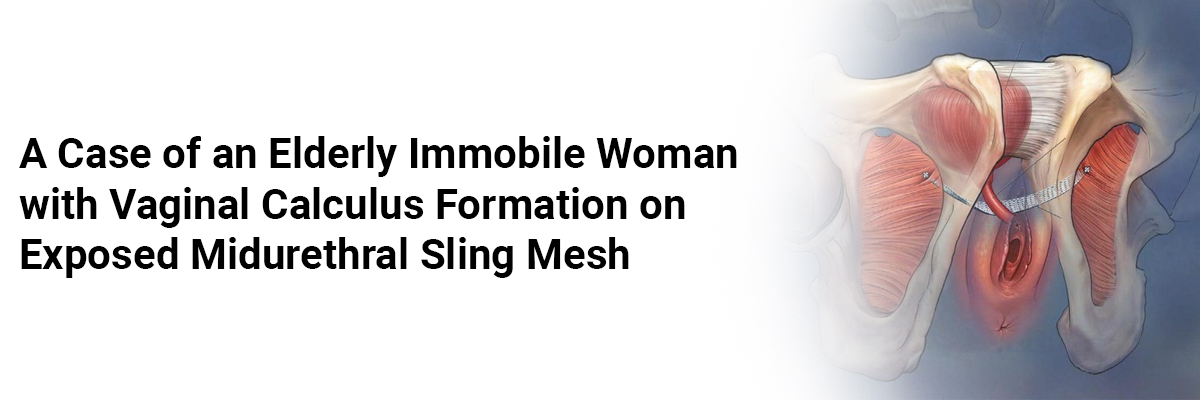
A Case of an Elderly Immobile Woman with Vaginal Calculus Formation on Exposed Midurethral Sling Mesh
The patient, a 74-year-old G4P4 female, reported new-onset postmenopausal vaginal bleeding characterized by the passage of multiple small clots and sudden vaginal pressure.
Her medical history included a total vaginal hysterectomy and midurethral sling surgery performed 5-7 years before her presentation for postmenopausal bleeding and stress urinary incontinence. She had had four prior vaginal births and was morbidly obese with a BMI of 71.97 kg/m2. She suffered from atrial fibrillation – was on apixaban; hyperlipidemia; had a history of breast cancer with unilateral mastectomy; hypothyroidism; pulmonary hypertension; chronic diastolic congestive heart failure; depression; stage 3b kidney failure; and lymphedema.
The initial pelvic exam, limited by her body habitus and mobility, revealed a grey-white, rough mass at the vaginal introitus, firm on bimanual examination, along with a malodorous bloody discharge.
Laboratory tests indicated leukocytosis without anemia. An abdominal CT scan suggested a 14 × 11 cm pelvic mass and mild bilateral hydronephrosis – later identified as a distended bladder due to hemorrhagic cystitis. The CT scan also showed a calcified mass in the vaginal or bladder area.
The patient underwent a pelvic exam under general anesthesia with cystourethroscopy and resection of the vaginal mass. Intraoperatively, gynecologic oncology and urology were consulted due to concerns about malignancy and potential urethral/bladder involvement. The calcified vaginal mass, adherent to the anterior vaginal wall, was excised at the midurethra. Underlying the calcified mass was exposed mesh from her previous midurethral sling, a small portion of which was removed, and the surrounding mucosa was sutured with delayed absorbable sutures. Cystourethroscopy showed no fistula or injury to the urethra or bladder but revealed gross bladder mucosal inflammation consistent with hemorrhagic cystitis. Clots and blood were evacuated from the bladder.
Surgical pathology identified the vaginal mass as a 2.4 × 2.0 × 1.7 cm white/tan, firm, calcified mass with visible mesh, consisting of amorphous material and calcifications without viable epithelium.
The lady developed an intraoperative fever and was started on broad-spectrum antibiotics, later transitioned to IV ceftriaxone until discharge. She had no additional fevers during her hospital stay. Blood and urine cultures were positive for Proteus mirabilis.
Her Foley catheter was removed on hospital day 6, and she was discharged to an extended care facility on oral cefuroxime. Follow-up with her cardiologist a month later indicated no recurrence of vaginal bleeding.
This case highlights the rare complication of large vaginal calculus formation on exposed midurethral sling mesh in an elderly immobile patient. The etiology is multifactorial, involving mesh erosion, chronic urinary incontinence, urinary stasis due to immobility and body habitus, lack of gynecological follow-up, and acute hemorrhagic cystitis secondary to Proteus mirabilis infection.
Source:Langer AJ, Saeed Z, Barrett E, et al. Case Reports in Obstetrics and Gynecology. 2024;2024(1):8287400.


Please login to comment on this article