 IJCP Editorial Team
A Rare Presentation of Dual Ovarian Pathologies
A 26-year-old female was diagnosed with a with stage IC well-differentiated mucinous ovarian cancer. The lady had a family history of breast cancer.
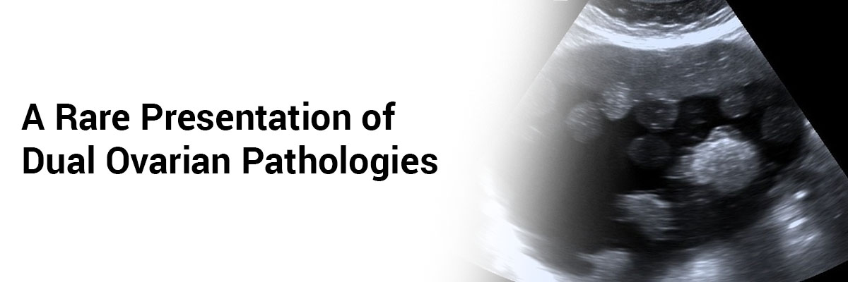
The prior diagnosis had been incidental – during the assessment and investigation for abnormal facial hair. Ultrasound (USG) abdomen and pelvis had shown a large cyst in the left ovary––20 cm––invading the abdominal space.
CT scan showed a large left complicated ovarian cyst with minimal obstructive ureter. Her CA-125 concentration was 22.3 U/mL. A laparoscopic left ovarian cystectomy was performed. Histopathological analysis revealed a well-differentiated ovarian mucinous carcinoma.
Two months later, the patient presented with progressively increasing abdominal distension. At this point, her CA-125 was 183 U/mL. USG abdomen, pelvis indicated gross ascites. Peritoneal fluid cytology indicated malignancy. CT scan revealed omentoperitoneal disease with large volume ascites.
The treatment plan included neoadjuvant chemotherapy. The radiological resolution was satisfactory after five cycles of carboplatin, paclitaxel, and bevacizumab. Thereafter, excision of the residual tumor was undertaken. Histology showed small cell carcinoma in the left ovary and a residual mucinous ovarian carcinoma. The right ovary had surface deposits of small cell carcinoma. A positron emission tomography scan (PETS) was done four weeks after surgery which showed progressive disease with ascites, along with an omental and peritoneal disease. The patient was started on cisplatin and etoposide chemotherapy. Only after one cycle as her health deteriorated and unfortunately, she could not survive.

IJCP Editorial Team
Comprising seasoned professionals and experts from the medical field, the IJCP editorial team is dedicated to delivering timely and accurate content and thriving to provide attention-grabbing information for the readers. What sets them apart are their diverse expertise, spanning academia, research, and clinical practice, and their dedication to upholding the highest standards of quality and integrity. With a wealth of experience and a commitment to excellence, the IJCP editorial team strives to provide valuable perspectives, the latest trends, and in-depth analyses across various medical domains, all in a way that keeps you interested and engaged.


Please login to comment on this article