
 IJCP Editorial Team
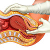
A simplistic approach to fibroids is outdated
The advancements in genomic sequencing and the regular emergence of new information forbid taking a simplistic approach to fibroids.
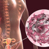
Fibroids affect nearly 80% of Black women and almost 70% of White women by the age of 50. Despite their prevalence, their exact molecular mechanisms remain unknown. However, genome-wide sequencing has helped in learning about the genetic alterations, both acquired and inherited, that facilitate the development of fibroids.
The commoner variant of fibroid has been linked to somatic mutations within them compelling tumor evolution and growth. Via Genomic sequencing changes in genes like MED12 and HMGA2, have been observed in 80%–90% of the cases.
Also the different phenotype in this variant may be due to the Genetic, environmental, and epigenetic factors. Proteogenomic analysis has suggested discrepancies in the molecular underpinnings between races.
A single nucleotide polymorphism, rs57542984, has been associated with maximal fibroid dimension, while two other single nucleotide polymorphisms have been linked to fibroid volume, rs10024805, and rs6605005.
The second category of fibroids is syndromic fibroids, resulting from germline mutations or heritable changes in DNA. Hereditary leiomyomatosis and renal cell cancer (HLRCC) is the most acknowledged genetic syndrome associated with fibroids, caused due to a heterozygous germline mutation in the fumarate hydratase (FH) gene.
Clinicians must recognize the heritable causes of fibroids. Early diagnosis is crucial for timely screening for cancer in patients and their affected family members. A thorough history and physical examination are keys to facilitating the diagnosis of HLRCC.
Fibroids from patients with HLRCC also show morphological features like increased cellularity, prominent eosinophilic nucleoli with perinucleolar halos, hyaline globules, and staghorn vasculature. Regarding immunohistochemistry, FH staining and 2-SC marker must be utilized to diagnose HLRCC.
Imaging of HLRCC fibroids may display a hyperintense signal on T2-weighted magnetic resonance imaging, which can aid in differentiating between HLRCC-caused tumors and common fibroids in a single scan.
An amalgamation of all these modalities will aid in affirming a clinical suspicion and prompt further genetic testing. Furthermore, a greater understanding of the genetic influences on the development and growth of fibroids will help in developing more targeted therapies in the future.
SOURCE- Volovsky M, Segars J. A simplistic approach to fibroids is outdated. F S Rep. 2021;3(1):11-12. Published 2021 Dec 7. doi:10.1016/j.xfre.2021.12.001
IJCP Editorial Team
Comprising seasoned professionals and experts from the medical field, the IJCP editorial team is dedicated to delivering timely and accurate content and thriving to provide attention-grabbing information for the readers. What sets them apart are their diverse expertise, spanning academia, research, and clinical practice, and their dedication to upholding the highest standards of quality and integrity. With a wealth of experience and a commitment to excellence, the IJCP editorial team strives to provide valuable perspectives, the latest trends, and in-depth analyses across various medical domains, all in a way that keeps you interested and engaged.
More FAQs by IJCP Editorial Team


Recent FAQs

Related FAQs


Medtalks is India's fastest growing Healthcare Learning and Patient Education Platform designed and developed to help doctors and other medical professionals to cater educational and training needs and to discover, discuss and learn the latest and best practices across 100+ medical specialties. Also find India Healthcare Latest Health News & Updates on the India Healthcare at Medtalks
Please login to comment on this article