 IJCP Editorial Team
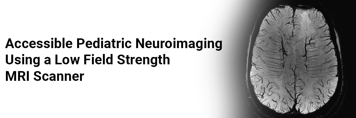
Accessible pediatric neuroimaging using a low field strength MRI scanner
Magnetic resonance imaging (MRI) has played an integral role in characterizing patterns of structural, functional, and metabolic brain development. Additionally, studies have demonstrated the influence of a child's physical and psychosocial environment on developing brain structure and function. A rate-limiting intricacy in these studies, however, is the high cost and infrastructural requirements of modern MRI systems. High costs mean many neuroimaging studies typically contain fewer than 100 people and are executed predominately in high-resource hospitals and university settings within high-income countries (HICs). As an outcome, our understanding of brain development, specifically in children residing in lower and middle-income countries (LMICs) is relatively narrow.
Low field systems, with magnetic fields of less than 100mT promise lower scanning costs and widespread international adoption, but routine low field pediatric neuroimaging has yet to be demonstrated. A recent study presented the first pediatric MRI data accumulated on a low-cost and assessable 64mT scanner in children falling between 6 weeks and 16 years of age group and reproduced brain volume estimates and developmental trajectories derived from 3T MRI data.
Although preliminary, these outcomes explain the potential of low field imaging as a possible complement to more conventional high field imaging systems and one that may further improve our knowledge of neurodevelopment in LMICs where malnutrition, psychosocial adversities, and other environmental exposures may significantly affect brain maturation.
Source: Neuroimage. 2021 Sep;238:118273. doi: 10.1016/j.neuroimage.2021.118273. Epub 2021 Jun 17. PMID: 34146712.

IJCP Editorial Team
Comprising seasoned professionals and experts from the medical field, the IJCP editorial team is dedicated to delivering timely and accurate content and thriving to provide attention-grabbing information for the readers. What sets them apart are their diverse expertise, spanning academia, research, and clinical practice, and their dedication to upholding the highest standards of quality and integrity. With a wealth of experience and a commitment to excellence, the IJCP editorial team strives to provide valuable perspectives, the latest trends, and in-depth analyses across various medical domains, all in a way that keeps you interested and engaged.


Please login to comment on this article