 IJCP Editorial Team
Clear Cell Carcinoma of the Cervix with OHVIRA Syndrome: A Rare Case Report
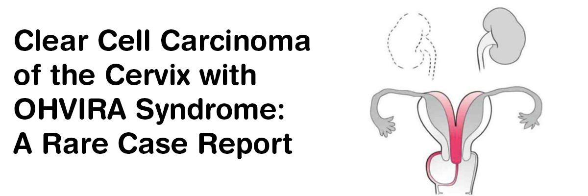
A report describes a case of a 52-year-old nulligravida woman who complained of intermittent vaginal spotting for 1 month, 2 years after menopause. She denied a history of smoking or medical diseases. She had no history of in-utero diethylstilbestrol (DES) exposure. Her family history contained an uncle who died of gastric cancer. She reported a cervical smear examination 11 months ago which was unremarkable.
Her Speculum examination was carried out that showed a single cervix along with a 2-cm mass on the cervix, which directed macrocarcinoma of the cervix. No parametrial or vaginal involvement was found. A biopsy taken from the tumor on the uterine cervix revealed CCC, comprising of atypical cells displaying enlarged nuclei with prominent nucleoli and clear-to-eosinophilic cytoplasm; the cells also displayed papillary structures with a hobnail-like appearance. Immunohistochemical staining demonstrated tumor cells to be positive for HNF-1β and napsin A. A negative Laboratory data was obtained concerning the patient’s squamous cell carcinoma antigens, carbohydrate antigen (CA) 125 levels, and CA19-9 levels.
Magnetic resonance imaging (MRI) of her pelvis demonstrated uterine didelphys and a mass measuring 1.5 × 2.0 cm located in the left endocervix not invading the parametria, adjacent organs, or regional lymph nodes. Also, a left-sided obstructed hemivagina was observed.
No evidence of lymph node metastasis or distant metastasis was observed in an enhanced computed tomography (CT) scan of the chest and abdomen, however, left renal agenesis was observed.
Concerning the findings of left renal agenesis, uterine didelphys, and obstructive hemivagina, she was diagnosed with OHVIRA syndrome and CCC of the cervix and classified as International Federation of Gynecology and Obstetrics stage IB1.
An abdominal radical hysterectomy was performed, including bilateral salpingo-oophorectomy and dissection of the bilateral pelvic lymph nodes. During the surgery, obvious effusion or metastasis was not observed in the abdominal cavity. Two corpora of the uterus were observed in her pelvis. Her left ureter led to the left cervix, and the renal side of the left ureter ended at the bifurcation of the common iliac vessels; thus, her left ureter was ectopic ureter, which was resected along with her left cervix and vaginal wall.
A macroscopic examination of the excised specimens revealed the presence of two cervixes, two corpora of the uterus, and a tumor measuring 1.0 × 2.0 cm on the left cervix. Her left ectopic ureter was guided to the left cervix and opened in the endometrium.
She got discharged postoperative day 10 after an uneventful recovery. Following surgery, she received no treatment and had no evidence of disease 12 months after therapy completion. Her postoperative course was uneventful.
Postoperative histopathology demonstrated uterine cervical CCC, without invasion of the uterine wall or lymph vessel and negative surgical margins. No metastatic lymph nodes on the 22 lymph nodes or invasion of the parametrium were observed, prompting a tumor-node metastasis classification of pT1b1N0M0. The histopathological findings demonstrated papillary growth and a tubulo-cystic growth pattern, which revealed CCC. Immunohistochemical staining displayed tumor cells to be positive for napsin A and HNF-1β, excluding block positivity of p16. Also, the loss of PAX2 expression, retained ARID1A and PTEN expression, and wild-type pattern of p53 staining was observed.
SOURCE- Tanase Y, Yoshida H, Naka T, et al. Clear Cell Carcinoma of the Cervix With OHVIRA Syndrome: A Rare Case Report. World J Oncol. 2021;12(1):34-38. doi:10.14740/wjon1362

IJCP Editorial Team
Comprising seasoned professionals and experts from the medical field, the IJCP editorial team is dedicated to delivering timely and accurate content and thriving to provide attention-grabbing information for the readers. What sets them apart are their diverse expertise, spanning academia, research, and clinical practice, and their dedication to upholding the highest standards of quality and integrity. With a wealth of experience and a commitment to excellence, the IJCP editorial team strives to provide valuable perspectives, the latest trends, and in-depth analyses across various medical domains, all in a way that keeps you interested and engaged.
More FAQs by IJCP Editorial Team
akang69
akang69
slot pulsa
slot pulsa
https://chateraise.org/menu/
slot gacor
kanjeng69
https://www.seansprimedining.com/dine
slot gacor
slot demo
akang69
slot gacor
slot pulsa
slot pulsa
akang69
https://valentinesdaycountdown.com/
slot jepang
slot gacor
slot gacor
slot88
slot gacor
slot gacor
akang69
akang69
akang69
akang69
slot gacor
slot gacor
slot gacor
https://www.alpinecafeandbakery.com/menu.html
https://applebeesmenu.us/applebees-lunch-menu/
https://bakeryandsweets-fest.com/product-highlight/
https://sevenseassushi.com/menu/
slot gacor
slot777
https://freakout.club/
slot gacor
slot gacor
https://rsgm.moestopo.ac.id/instalasi-rawat-jalan/
slot gacor
slot pulsa
slot gacor
slot gacor
http://www.motohom.co.in/
slot pulsa
https://intervencion.uahurtado.cl/
slot pulsa
slot pulsa
https://www.kp2.it.maranatha.edu/
slot gacor
slot gacor
mahjong ways
slot gacor
https://azure3.test.utah.edu/
https://iceam.unimap.edu.my/
https://sindika.co.id/contact/
slot gacor
slot gacor
slot pulsa
https://jhep.unimap.edu.my/
slot gacor
Kartu Pokémon Naik Harga!! Penghobi Naik Drastis, Bermain No Limit Dapat Cuan Berlimpah
Resep Putar Hemat WD Paus di Sugar Rush, Murah Namun Ampuh!
Seseorang Terlihat Jauh Lebih Muda dari Usianya, 7 Game Big Bass Crash yang Menguntungkan
Tukang Bubur Demi Beli Honda Megapro Terbaru, Main PGSoft di Kanjeng Jadi Jutawan!
slot gacor
slot pulsa
https://portal.kaafuni.edu.gh/
https://www.thedeenshow.com/
https://elektro.trunojoyo.ac.id/
https://pgsd.trunojoyo.ac.id/
http://www.gmci.in/
https://www.medigunakhisar.com/contact/
https://www.medigunakhisar.com/category/doktorlar/
https://www.medigunakhisar.com/about/
https://www.oscarstores.com/
https://ritalabailaora.com/
https://journal.stiepertiba.ac.id/official/
https://shopifyvps.trackship.co/
https://krandeganbayan.id/
https://www.imik.edu.in/
https://www.natasshaselvaraj.com/
http://ebphtb.banjarnegarakab.go.id/
https://bigbiteonpitt.com.au/
https://sigen.kaltimprov.go.id/
http://rju.parco.gov.ba/
https://samajpragatisahayog.org/
https://bendismea.be.gov.ng/
https://mozaiktravel.id/
https://xm42newdev.wpengine.com/
https://epr.rw/
https://tribelio.page/slotpulsa
https://mhs.akpertgkfakinah.ac.id/
http://lms6.digivarsity.com/
https://ssr.vinayakamission.com/
https://prelnor.molg.go.ug/gallery/
https://englishfocus.upstegal.ac.id/
akang69
https://virtex.canadianminingexpo.com/
slot pulsa
slot bet 200
slot online
convocation.aiou.edu.pk/
https://qna.bpsaceh.com/
https://storage.therapyrooms.com/
https://www.inaexport.id/
https://vmrfdu.edu.in/
https://www.jniemann.it/
https://katalog.intanonline.com/view/
akang69
akang69
akang69
https://affordableadsgroup.com/
https://doktor.fisip.hangtuah.ac.id/
https://um.originmena.com/
https://primemarkets.com.sa/branches
https://primemarkets.com.sa/
https://www.aqtiverse.in/images/
slot gacor
https://rokomarifood.com/
https://beritrust.com/
https://dispora.sumbarprov.go.id/imgs/
slot pulsa
http://pubma.edostate.gov.ng/
https://palermo.if.ua/
akang69
slot qris
http://husc.hueuni.edu.vn/
https://www.grupogasca.com/
slot pulsa
https://ukm.stiedharmaputra-smg.ac.id/
https://ppid.mukomukokab.go.id/
slot gacor
https://s1cs.stmikroyal.ac.id/
https://terlaksana.co.id/img/
slot pulsa
https://graduation.sjctni.edu/SJCGRAD24/abc_id.php
https://admission.sjctni.edu/
https://camarabonitodeminas.mg.gov.br/oficios/
https://kmob.jabarprov.go.id/
https://ssm.school.ssmetrust.in/
https://cjnc.ppnijateng.org/
https://paisaje.age-geografia.es/
slot pulsa
hasilwin
demototo
http://majaslapa.lv/media/
slot gacor
https://sumpah.ppnijateng.org/
https://www.uniabuja.edu.ng/
https://training.super5.org/
slot pulsa
slot gacor
https://mmi.edu.pk/find-a-doctor/
https://lonsuit.unismuhluwuk.ac.id/
slot pulsa
slot gacor
https://taxicarudaipur.com/
slot pulsa
slot gacor
slot gacor
slot gacor
slot pulsa
https://siraberu.mixh.jp/
slot gacor
slot pulsa
slot gacor
https://hkg.methodist.org.hk/
slot gacor
https://www.asopa.org/
https://fisip.unismuhluwuk.ac.id/
slot gacor
https://www.panganku.org/id-ID/semua_nutrisi
https://ejurnal.ikippgribojonegoro.ac.id/
slot gacor
https://katalog.intanonline.com/view/
https://stikesgrahaedukasi.ac.id/
https://kec.slogohimo.wonogirikab.go.id/
https://www.facesulavirtual.net/efaces/
https://www.gratisongkir.id/
https://bukupdpi.klikpdpi.com/
https://ilp.mizoram.gov.in/
https://www.campdoha.org/
slot gacor
slot gacor
https://katalog.intanonline.com/
slot gacor
https://munabarat.go.id/index.php
https://youtube.klikpdpi.com/
https://katalog.intanonline.com/lazada.IM.im-msgbox
https://understandquran.com/
https://youtube.klikpdpi.com/
https://gratisongkir.id/console/
https://gratisongkir.id/vagrant/
https://www.gratisongkir.id/uploads/
https://www.gratisongkir.id/assets/
https://snyderfamilyband.com/
https://handholding.mizoram.gov.in/
https://dconline.mizoram.gov.in/
https://siva.umkendari.ac.id/
https://www.gthlcanada.com/
https://www.gratisongkir.id/assets/
https://www.vidyasthalilawcollege.com/
togel toto
slot gacor
https://www.facesulavirtual.net/
hasilwin
https://amertamedia.co.id/
https://www.gulfstarsauto.com/
https://aptika.id/
https://www.bytestechnolab.com/
slot gacor
slot deposit 10k
slot singapore
slot thailand
AKANG69
AKANG69
https://snyderfamilyband.com/
slot gacor
https://epaper.notunshomoy.com/
https://development.upr.ac.id/
https://fornas.kebijakankesehatanindonesia.net/
slot gacor
slot pulsa
https://e-anatolh.com/
slot gacor
slot thailand
slot pulsa
kanjeng69
https://loudhelp.com/
slot thailand
https://ipss-addis.org/
slot gacor
https://engineering.upstegal.ac.id/
slot gacor
https://sipaksi.unpak.ac.id/
https://pbi.teknik.unmuha.ac.id/
https://bkd.inkhas.ac.id/files/
https://barcode.akbidyahmi.ac.id/
https://sdcirebonislamic.cerdig.com/
https://tilganga.org/
https://siperjaka.ms-langsa.go.id/
slot pulsa
https://dinsos.bekasikab.go.id/
https://stkq.alhikamdepok.ac.id/
http://fmipa.upr.ac.id/
https://beasiswaperintis.id/
slot pulsa
slot pulsa
https://www.thebestflushingtoilet.com/
https://sdiassalaftahfidzulquran.cerdig.com/
https://sv-388.asia/
https://lppm.upr.ac.id/
https://peraturan.upr.ac.id/
https://simpeg-tb.id/
https://contentacademy.id/
https://ws-168.org/
slot pulsa
https://simpeg-tb.id/
https://www.lagrangepointsbrussels.com/
https://admisi.uinsaid.ac.id/img/
slot gatot kaca
https://www.multimarcasgrupovianorte.com.br/
https://silajara.kepulauanselayarkab.go.id/
slot pulsa
https://bapenda.batubarakab.go.id/run/
https://dosen.billfath.ac.id/sites/
gatot kaca slot
http://sibkd.semarangkab.go.id/ekgb/temp/
http://jak.faperta.unand.ac.id/sites/
slot pulsa
slot kamboja
https://dinsos.bekasikab.go.id/yasha/
http://agribisnis.faperta.unand.ac.id/
https://hondayogyakarta.id/
http://jak.faperta.unand.ac.id/sites/
https://dinsos.bekasikab.go.id/hunt/
https://dinsos.bekasikab.go.id/dagger/
http://tanah.faperta.unand.ac.id/potion/
https://dinsos.bekasikab.go.id/dagon/
https://dinsos.bekasikab.go.id/potion/
https://tourism.lgu-santol.gov.ph/
https://fdik.uinmataram.ac.id/rune/
https://v2.poltekpelni.ac.id/fiend/
https://simbada.kalbarprov.go.id/
https://slotpulsa2025.net/
https://slotindosat.net/
https://www.scootersforknee.com/
http://sibkd.semarangkab.go.id/sib/excel/
https://www.worldminner.com/
slot gacor
https://literate.nusaputra.ac.id/lib/
https://potomacofficersclub.com/wp-content/cache/
https://literate.nusaputra.ac.id/docs/
https://literate.nusaputra.ac.id/api/
https://www.idecesar.gov.co/images/
http://sim-epk.poltekkes-medan.ac.id/
http://sibkd.semarangkab.go.id/kartukorpri/pages/
http://sibkd.semarangkab.go.id/kartukorpri/pages/
https://buycapybara.com/
https://desasungaibuluhungar.id/
https://pgmi.unupurwokerto.ac.id/slotzeus/
https://pgmi.unupurwokerto.ac.id/slotthailand/
slot dana
https://admissions.kmtc.ac.ke/
https://permana.upstegal.ac.id/run/
https://permana.upstegal.ac.id/pages/run/
https://stkq2.alhikamdepok.ac.id/
slot pulsa
https://www.allnursejobdescriptions.com/
https://mbkm.uim.ac.id/wp-includes/images/
https://tinjukosimaling.baritotimurkab.go.id/
https://www.silenteye.org/
slot pulsa
https://upscpdf.com/img/
https://keusatker.badilag.net/file_import/
https://bisnis.poltekkesdepkes-sby.ac.id/wp-content/
Slot Bca
Slot Qris
Scatter Hitam
Slot Thailand
Slot Qris
Slot Thailand
Slot gacor
Slot pulsa
Slot zeus
Slot zeus
https://bisnis.poltekkesdepkes-sby.ac.id/wp-content/
https://gresikunited.com/run/
slot gacor
https://pusppm.poltekkesdepkes-sby.ac.id/file/
https://gameplayterbaru.com/
deposit pulsa indosat
https://jdih.tubankab.go.id/js/
https://jdih.tubankab.go.id/library/
https://jdih.tubankab.go.id/img/
https://jdih.tubankab.go.id/gallery/
slot thailand
https://epanel.cblu.ac.in/-/
http://bjkang14.postech.ac.kr/wordpress/
http://bjkang14.postech.ac.kr/wp-includes/assets/
http://bjkang14.postech.ac.kr/wp-includes/images/
https://arktisdesign.com/
https://www.allnursejobdescriptions.com/
https://ziare-online.com/
slot gacor
https://e-jazirah.com/
slot qris
https://www.ultimatepethub.com/
https://slotovo2025.com/
https://www.mobeyday.com/
https://staipati.ac.id/
scatter hitam
slot thailand
scatter hitam
akang69
akang69
akang69
slot qris
slot dana
mahjong ways 3
mahjong wins 3
https://assignmenthacks.com/
Slot Pulsa
Slot pulsa
Slot pulsa
https://acehpos.id/
https://archerysportsindia.com/
https://mrsavnpolytechnic.com/
https://nandanavanamorganics.com/
https://leeupvcsolutions.com/
https://lelaskinclinic.com/
slot thailand
https://thebharatschool.com/
https://mealsandmilemarkers.com/
https://ecustomssecure.com/
https://wallstreetenglish.edu.mm/
https://audio-constructor.com/
https://www.pulsaaxis.net/
https://www.akangseru.com/
https://ukrainemarriageguide.com/
akang69
https://www.akangseru.com/
https://catalinaislandbrewhouse.com/
slot shopeepay
slot shopeepay
10 situs togel
https://captionstore.com/
https://gettranslate.org/
https://terrariumtvultimate.com/
https://www.pryorok.com/
https://saralgyaan.com/
https://ginghameats.com/
slot shopeepay
https://gettranslate.org/
https://captionstore.com
https://enjoyislamabad.com
https://waveites.com
https://voicenusantara.id/
https://mmabreakdown.com/
https://bobmccoyart.com/
https://perpusdajawatengah.id/
https://ziggyoficialbr.com/
slot pulsa
https://linkr.bio/akang69/
https://dev.cfar.org/
https://saveursdailleurs.net/
https://joy.link/akang69
https://ponpesmodernal-imantebelian.ponpes.id/sites/
http://themockingbirdkc.com
http://akang699.kesug.com/
http://akang69.rf.gd/
https://www.ongrace.com/
https://my-home-zen-spa.com/
https://cebofil.org/
http://ironwooddesignstudio.com
https://cebofil.org/
https://pulsa-akang69.tumblr.com/
https://bandar-akang69.tumblr.com/
https://akang69daftar.tumblr.com/
https://sikelor.parigimoutongkab.go.id/vendor/agen-pulsa/
https://www.deviantart.com/slot-gacor-akang69/about
https://www.yuswohady.com/
https://indonesiaindustryoutlook.com/
https://bikeindex.org/users/akang69
https://rivielle.com/
https://issuu.com/akang69
https://heylink.me/akang69z
https://graphicography.id/slotbca/
http://oyannews.com/az/slotdemo/
https://www.laser-travel.com.np/
https://sim-epk.poltekkesjogja.ac.id/slotpulsa/
https://uddoktahoi.com/
http://www.lightcodes.net/
https://wave-gate.lightcodes.net/
https://pohwates-bjn.desa.id/slotdepo/
https://zabava.bluespot.hr/
https://bluespot.hr/
https://www.thethingsnetwork.org/u/akang69/
https://bikeindex.org/users/akang69/
https://www.masterassignment.com/
https://getyourexamdone.com/
https://www.penzion-miromar.cz/
https://sardogs.net/
https://king-lbent.com/
https://lamstyle.com/
https://pulsaindosat.us.com/
https://pulsaindosat.eu.com/
https://pulsaindosat.uk.com/
https://tamilmv.ws/
https://slotindosatt.com/


Please login to comment on this article