Ileo-ileal Knotting – A Case Report
A
19-month-old boy presented with multiple vomiting episodes. His body weight was
9.3 kg, and his height was 80 cm.
Initial
clinical examination revealed irritability and signs of dehydration.
The child
was immediately started on dehydration correction; however, he experienced
multiple episodes of hematemesis within four hours of admission.
Upon
reassessment, the child was drowsy and exhibited signs of compensated shock,
including cold extremities, weak peripheral pulses, and a prolonged capillary
refill time, with normal age-appropriate blood pressure. His abdomen was
distended, with absent bowel sounds, guarding, and rigidity.
Six hours
post-admission, his hemoglobin levels dropped from 9.2 g/dL to 6.9 g/dL –
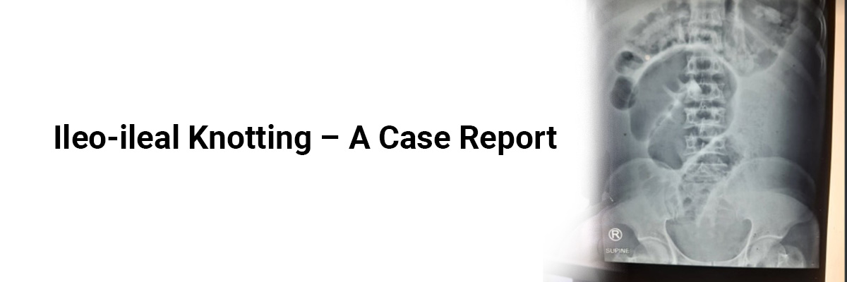
necessitating a transfusion of packed red blood cells. An abdominal X-ray
revealed multiple air-fluid levels indicative of intestinal obstruction.
Computed tomography (CT) of the abdomen showed dilated small bowel loops with
intramural air, consistent with early small bowel ischemia. The child's
Pediatric Risk of Mortality (PRISM) score was 19.
An
emergency exploratory laparotomy uncovered ileo-ileal knotting, with a
gangrenous ileum segment approximately 50 cm long, extending to one foot
proximal to the ileocecal junction.
The
gangrenous ileum was resected, and gut continuity was restored via
end-to-end anastomosis. The immediate postoperative period was uneventful.
On the
sixth postoperative day, after initiating feeds, the child developed chylous
ascites, indicated by a whitish peritoneal drain collection, with serum amylase
at 264 IU/L, serum lipase at 365 IU/L, and drain fluid triglycerides at 277
mg/dL. The child was started on an octreotide infusion and a fat-free diet.
Serial ultrasound monitoring and abdominal girth measurements showed no further
collection.
Octreotide
was gradually tapered off, and feeds were reintroduced. The child was
discharged on the seventeenth postoperative day without gastrointestinal
symptoms or complications during the nine-month follow-up.
In
ileo-ileal knotting, one ileal loop remains static while another encircles it,
forming a knot. The condition has been associated with the appendix or Meckel's
diverticulum. The knot formation initiates a cycle of intestinal occlusion and
ischemia due to continuous peristalsis and vascular pulsations, leading to
gangrene. Untying the knot suffices if all segments are viable, as recurrence
is rare. For irreversible ischemia, controlled decompression with a needle or
enterotomy should precede the resection of congested segments.
Factors such as a freely mobile small intestine and redundant sigmoid colon with a long, narrow mesentery are implicated in ileo-sigmoid knotting. Treatment involves early, aggressive IV fluid resuscitation, nasogastric tube insertion, and broad-spectrum IV antibiotics. Once stabilized, an emergency laparotomy is performed. Although rare, ileo-ileal knotting should be considered in differential diagnoses of small bowel obstruction.
Source: Harini JJ, Satish JK, Rajeswari PA, Prabhu GG.
Indian Pediatrics.2024:S097475591600587.


Please login to comment on this article