 IJCP Editorial Team
Intramural pregnancy: A case report.
A case report describes a 24-year-old patient with a medical history of one early pregnancy miscarriage not requiring surgical intervention, bronchial asthma, animal hair allergy, foot surgery, and mild obesity. For her asthma, she used beclometasone dipropionate/ formoterol fumarate inhaler on demand. She was 8 + 4 weeks pregnant and had a history of cesarean section 6 months back due to obstructed labor. She did not have any fertility treatment, never used an intrauterine contraceptive device and had no history of pelvic inflammatory disease. She used condoms as a contraceptive method.
One month before the presentation, she had a positive home pregnancy test, which couldn't be visualized by ultrasound two weeks later. Subsequent ultrasound assessment after two weeks interpreted ectopic pregnancy with a suspicion of an intramural pregnancy and hence she was immediately admitted.
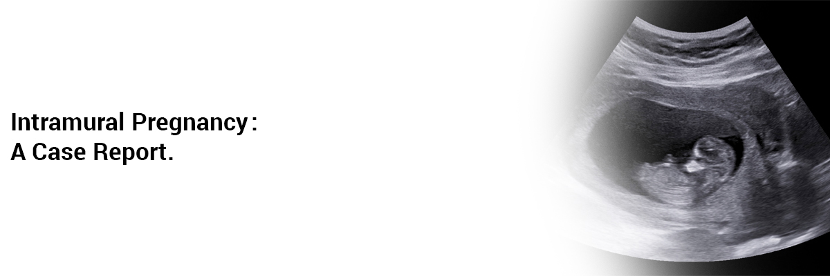
A moderately developed endometrium, inconspicuous adnexa on both sides, without free intraabdominal fluid were observed in an Ultrasound examination. Also, the left side of the uterus showed a 43x35mm mass with a gestational sac and a viable embryo (crown-rump length 21 mm), which did not communicate with the fallopian tube and uterine cavity.
βHCG was found to be 53,000 miU/ml, compatible with the calculated gestational age. Thus an intramural ectopic pregnancy (iEP) was suspected and treatment options were discussed with the patient.
Her history guided the operation to be primarily laparotomy, conceivably with further hysteroscopy and curettage.
Procedural revealed intramural pregnancy near the left tubal os, confirming the sonographic findings. The affected area was removed following the dissection and preservation of the tube.
Her postoperative period remained uneventful, except the βHCG levels dropped to 5800 mIE/ml for which she received gynaecological advice.
The operative findings got confirmed by the Histopathological examination.
Two and a half months following surgery, the patient presented with an intrauterine pregnancy, for which she was advised-
- Termination or
- Continuance of the pregnancy with close monitoring and premature delivery after 34 weeks of pregnancy.
SOURCE- Case Rep Womens Health. 2020;27:e00215. Published 2020 May 6. doi:10.1016/j.crwh.2020.e00215

IJCP Editorial Team
Comprising seasoned professionals and experts from the medical field, the IJCP editorial team is dedicated to delivering timely and accurate content and thriving to provide attention-grabbing information for the readers. What sets them apart are their diverse expertise, spanning academia, research, and clinical practice, and their dedication to upholding the highest standards of quality and integrity. With a wealth of experience and a commitment to excellence, the IJCP editorial team strives to provide valuable perspectives, the latest trends, and in-depth analyses across various medical domains, all in a way that keeps you interested and engaged.


Please login to comment on this article