 IJCP Editorial Team
Myxoid ovaries resembling malignancy
A report describes a case of a 12-year-old girl who previously received a provisional diagnosis of suspected appendicitis and an exploratory laparoscopic procedure that concluded no signs of infection of the appendix or other anatomical sites.
Further anamnesis and physical examination revealed pain in the abdomen for two months; without any enlargement in the abdomen or bleeding from the genitals. The patient had not menstruated, and her defecation and urination were normal.
Ultrasound examination showed anteflexi uterus (measuring 4.56 x 2 cm), with an end line (+). The appearance of a hypoechoic adnexa with a solid area (measuring 4.53 x 3.74 cm), ill-defined, vascular score 4, without acoustic shadow raised the suspicion for ovarian malignancy, hence a surgical procedure in the form of a conservative surgical staging Laparotomy was planned.
Intraoperatively, a mass was obtained from the right ovary. The patient's uterus and left ovary were within normal limits, with no lymph node enlargement and no masses in the colon, omentum, liver, ileum, or peritoneum.
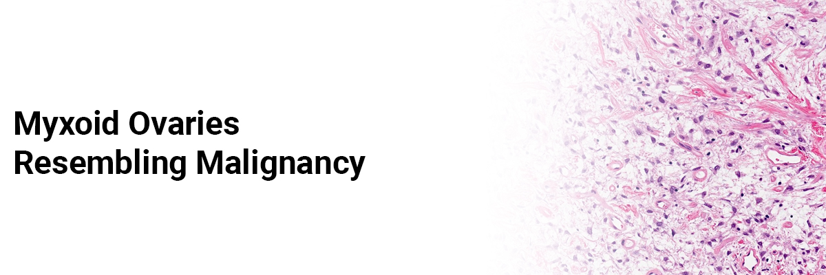
Microscopic examination of the ovarian and omental tissues showed connective tissue stroma containing proliferating cells with oval-spindle nuclei, hyperchromatic against a myxoid stroma background, and adipose tissue containing hyperemic capillaries. It also showed tubal tissue and its projections covered by cuboidal epithelium without any tumor cells.
Peritoneal fluid showed the microscopic distribution of mesothelial groups, lymphocytes, and some PMN leukocytes. It showed no malignant tumor cells.
The patient was finally diagnosed with Myxoma Uteri.
Ovarian myxoma is rare and difficult to diagnose because of the non-specific symptoms. Patients with Relatively younger ages are often misdiagnosed and thus must be differentiated from other ovarian lesions with myxoid changes. It's crucial to differentiate this tumor carefully from other benign and malignant myxoid lesions of the ovary because it resembles other forms of ovarian malignancy closely.
Fajriman F, Antonius PA, Muhammad S. Myxoid ovaries that resemble malignancy in young girls: a case report. AOJ. 2023;7(2). DOI: https://doi.org/10.25077/aoj.7.2.473-478.2023

IJCP Editorial Team
Comprising seasoned professionals and experts from the medical field, the IJCP editorial team is dedicated to delivering timely and accurate content and thriving to provide attention-grabbing information for the readers. What sets them apart are their diverse expertise, spanning academia, research, and clinical practice, and their dedication to upholding the highest standards of quality and integrity. With a wealth of experience and a commitment to excellence, the IJCP editorial team strives to provide valuable perspectives, the latest trends, and in-depth analyses across various medical domains, all in a way that keeps you interested and engaged.


Please login to comment on this article