 IJCP Editorial Team
Painful Abdominal Wall Mass Requiring Resection in the Second Trimester
A recent report describes a case of a 28-year-old female, gravida 2, para 1, who presented at seven-week gestation for routine antenatal care.
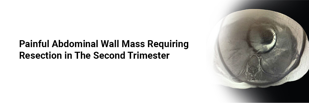
The patient reported noticing a lump in her abdomen near the umbilicus two months before becoming pregnant, which, during pregnancy, grew rapidly. Ultrasound and subsequent MRI revealed benign characteristics and no evidence of local or distant extension. She received a recommendation for expectant management during pregnancy and surgical excision after delivery. However, the continued growth and pain-directed surgical resection of mass (measuring 3.5 cm) at 25 weeks of pregnancy.
Histopathology showed bland spindle cells in long fascicles with compressed blood vessels, perivascular edema, and focal extravasated red blood cells. Immunohistochemical stains were positive for beta-catenin, smooth muscle actin (SMA), and desmin. Thus, she received a histological diagnosis of desmoid-type fibromatosis.
The patient showed an uneventful recovery and delivered vaginally at full term a female infant with normal Apgar scores.
A seven-month follow-up MRI after the resection showed no residual mass or recurrence. The management plan includes continuing with clinical and radiological surveillance.
Stemmer SM, Gomes C, Cardonick EH. A Case of Painful Growing Abdominal Wall Mass during Pregnancy Requiring Resection in the Second Trimester. Case Reports in Obstetrics and Gynecology. 2024;2024. https://doi.org/10.1155/2024/5881260

IJCP Editorial Team
Comprising seasoned professionals and experts from the medical field, the IJCP editorial team is dedicated to delivering timely and accurate content and thriving to provide attention-grabbing information for the readers. What sets them apart are their diverse expertise, spanning academia, research, and clinical practice, and their dedication to upholding the highest standards of quality and integrity. With a wealth of experience and a commitment to excellence, the IJCP editorial team strives to provide valuable perspectives, the latest trends, and in-depth analyses across various medical domains, all in a way that keeps you interested and engaged.


Please login to comment on this article