 IJCP Editorial Team
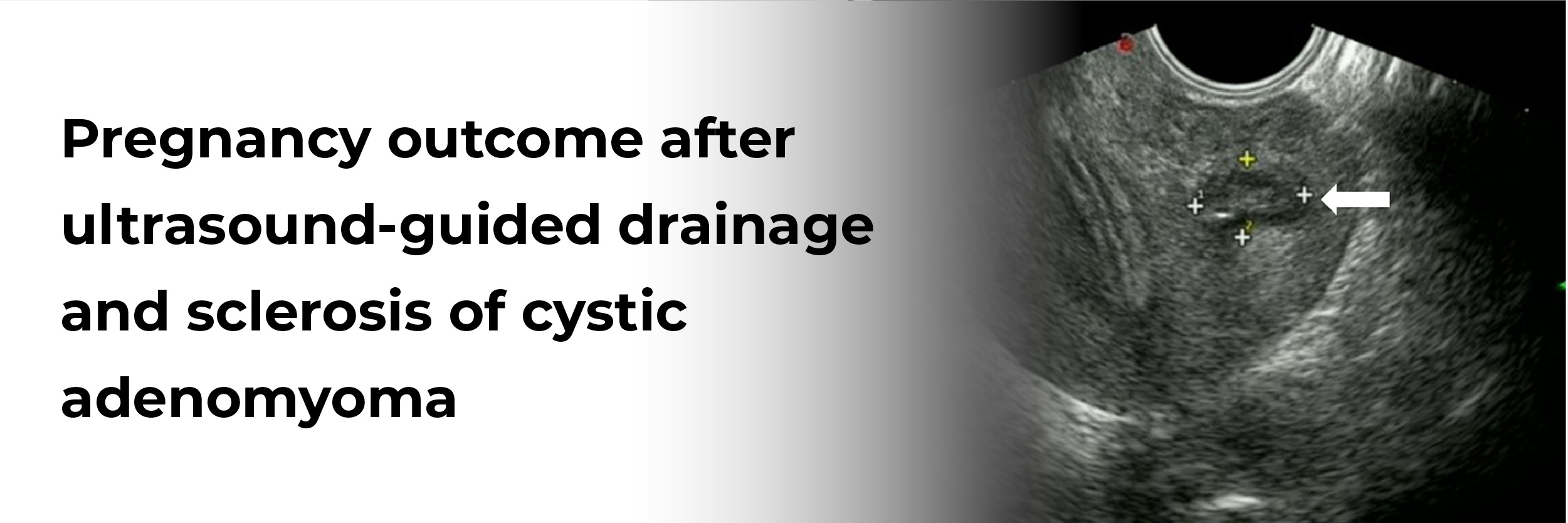
Pregnancy outcome after ultrasound-guided drainage and sclerosis of cystic adenomyoma
A report describes a case of a 22-year-old primiparous female with a past medical history significant for repaired Tetralogy of Fallot, asthma, bronchiectasis, and previously described cystic adenomyomas. Her past medical history revealed that she presented with bloating and constipation in 2014, and was discovered with an abdominal mass on a pelvic exam. Her ultrasound imaging at that time revealed a large simple ovarian cyst and she underwent a diagnostic laparoscopy with planned laparoscopic ovarian cystectomy, which showed a mass originating from the uterus and the procedure was then aborted. Following the procedure, she underwent an MRI which revealed a large myometrial cyst of size ~13.3 × 9.5 × 10.4 cm with an inferior locule of ~8.8 × 10.1 × 10.4 cm with simple appearing fluid and a superior locule of ~3.5 × 6.6 × 7.7cm with hemorrhagic material separated by thick septation. Imaging showed no evidence of congenital uterine anomaly, with a single endometrial cavity, and normal ovaries with small ovarian lesions suggestive of endometriomas. IR aspiration of the cyst was carried out with repeat imaging displaying a reduction in size to 9.4 × 9.1 × 9.4 cm, correlating with symptomatic relief.
Over the next three years, she experienced an intermittent recurrence of symptoms and imaging also demonstrated recurrence of myometrial cystic adenomas about 3–4 times in different locations of the uterus. One such episode was also associated with severe hydronephrosis from cyst compression of the ureter, which resolved after drainage with no long-term renal damage.
She experienced five additional IR aspirations over three years, with the use of ethanol sclerotic agents (10 to 200 mL) during three aspirations. In one aspiration 500 mg in 20 mL of doxycycline sclerosis was used. All of these procedures were uncomplicated.
She finally underwent medical management with GnRH agonist therapy for 3 months and transitioned to extended cycle oral contraceptives, and subsequently to an etonogestrel implant. Additionally, she was suggested surgical management with myometrial cystectomy or hysterectomy, however, she continued hormonal suppression with intermittent attempts at IR drainage as she desired fertility preservation.
A recent case report is a follow-up of the aforementioned patient during her pregnancy three years after treatment of myometrial cysts. She presented at 11 weeks and 1 day for a new prenatal visit.
She did not report any symptoms in the first and second trimesters of her pregnancy to suggest the growth of her myometrial cyst. Additionally, her first-trimester ultrasound did not show any myometrial cysts. Her anatomy scan at 20 weeks was significant for a placental lake, with the largest of size 2.26 × 4.55 × 4.02 cm with multiple other smaller 1 × 1 × 1 cm chorionic cysts which were visualized on the fetal aspect of the placenta. The placenta did not show any signs of a placenta accreta. Her complex history directed growth ultrasound at 32 weeks which revealed fetal growth restriction (FGR) with an estimated fetal weight (EFW) of 1590g (8th percentile) with normal umbilical artery dopplers.
She thereafter started appropriate antenatal testing which continued to be reassuring until term. A growth scan was repeated at 36 weeks which showed an EFW of 2204 g (5th percentile) and umbilical artery dopplers were again normal. She underwent scheduled induction of labor for FGR at 38 weeks, which was overall uncomplicated and led to an uneventful vaginal delivery of a 6 bs 13.7 oz (3.11 kg) baby boy with Apgars of 5, 9.
Duncan JM, Janssen M, Shlansky-Goldberg R, Hirshberg A, Neff P. Pregnancy outcome after ultrasound-guided drainage and sclerosis of cystic adenomyoma. J Case Rep Images Obstet Gynecol 2021;7:100097Z08JD2021.

IJCP Editorial Team
Comprising seasoned professionals and experts from the medical field, the IJCP editorial team is dedicated to delivering timely and accurate content and thriving to provide attention-grabbing information for the readers. What sets them apart are their diverse expertise, spanning academia, research, and clinical practice, and their dedication to upholding the highest standards of quality and integrity. With a wealth of experience and a commitment to excellence, the IJCP editorial team strives to provide valuable perspectives, the latest trends, and in-depth analyses across various medical domains, all in a way that keeps you interested and engaged.


Please login to comment on this article